Friday, 4 December 2015
INVERTEBRATES
They are mostly marine, few are found in fresh water.
Mostly are assymetrical animals, no definite shape to the body.
These are primitive animals, multicellular with cellular grade of organization.
Adult sponges are sessile, that is they need a substratum to attach themselves to a surface and do not move.
Due to the sessile nature, sponges are filter feeders.
Digestion is intracellular.
The body of sponges is supported by a skeleton made of spicules or spongin fibres.
Sexes are not separate, they are hermaphrodites. Hermaphroditism - condition where eggs and sperms are produced by the same individual.Sponges reproduces asexually by fragmentation and sexually by formation of gametes.
Fertilization is internal.
Indirect development, having a larval stage which is morphologically distinct from the adult.
Sponges have water transport system. Water enters through minute pores (ostia) in the body wall into a central cavity known as spongocoel. From the spongocoel water goes out through the osculum. This water system aids in food gathering, respiratory exchange and removal of wastes.
These are collar cells, they line the spongocoel and the canals.
Tuesday, 1 December 2015
INVERTEBRATES
Classification of Phylum Protozoa:-
Phylum Protozoa divided into two sub-phylums1. Sub Phylum Plasmodroma
2. Sub Phylum Ciliophora
1. Sub Phylum Plasmodroma
This sub phulum is further divided into 6 super classes.
1. Mastigophora
this divided into 2 classes
a. Phytomastigophora
b. Zoomastigophora
2. Opalinata
3.Sarcodina
this is also divided into 2 classes
a. Hydrailea
b. Autotractea
4.Apicomplexa
5.Mesospora or Myxospora
6. Microspora
2. Sub Phylum Ciliophora
This sub phylum divided into 1 super class.
Super class Ciliata
which is further divided into 3 classes
1. Kinetofragmenophora
2.Oligohymenophora
3. Polyhymenophora
Sunday, 29 November 2015
WHAT ARE INVERTEBRATES?
Invertebrates Classification
Some invertebrate phyla are:
Phylum Protozoa
Phylum Porifera (Sponges)
Phylum Cnidaria (Coelentrata)
Phylum Ctenophora :
Phylum Platyhelminthes
Phylum Aschelminthes (Nematoda)
Phylum Annelida (Segmented worms)
Phylum Arthropoda
Phylum Mollusca
Phylum Echinodermata
Phylum Protozoa
- Members of this phylum are commonly known as sponges.
- Habitat - They are mostly marine, few are found in fresh water.
- Body symmetry - mostly are assymetrical animals, no definite shape to the body.
- Level of organization - These are primitive animals, multicellular with cellular grade of organization.
- Digestion - Digestion is intracellular.
- Reproduction- Reproduction of both means sexually as well as asexual.
- Asexually by mean of binary fission,by budding etc.
- Sexually by mean of conjugation.
- Examples- Paramecium,Amoeba,plasmodium etc.
WHAT ARE INVERTEBRATES?
List of Invertebrates
Invertebrates are the most diverse organisms present on earth. Almost 95% of the animal populations are of the invertebrates. Based on the International Union regarding Conservation of Nature at the time of 2009 more than 1.3 million invertebrates were identified. Invertebrates make up around 75% of the recognized species on Planet.
The actual number of invertebrates is unknown, there are several predictions that there may be tens of millions of invertebrates, majority being the insects. New species of invertebrates are being discovered regularly, another worrying fact is that there is insufficient information about these organisms, the invertebrates might be going extinct and the scientists would never know that they even existed.
Below are a list of invertebrates -
Crustaceans, Centipedes, Ants, Wasps, Spiders, Locusts, Honey bees, Termites, Cockroach, Grasshoppers, Crickets, Stick insect, Mantis, Crabs, Sponges, Starfish, Unio, Leeches, Earthworms, etc.

WHAT ARE INVERTEBRATES?
Characteristics of Invertebrates
:-
General characteristics of invertebrates are as follows:
General characteristics of invertebrates are as follows:
- The main characteristic that separates invertebrates from other organisms is the absence of the spinal column and backbone.
- They are multicelluar organisms, they completely lack cell walls.
- They are devoid hard bony endoskeleton.
- Due to the lack of complex skeletal systems, some invertebrates tend to be slow and small in nature.
- Due to the lack of the backbone and complex nervous system the invertebrates cannot occupy mulitple environments, though they are found in the harshest of the environments.
- Invertebrates live all over the world in various habitats.
- Body is divided into three parts - head, thorax and the abdomen.
- They do not have lungs for respiration.
- Respiration is through skin.
- Some invertebrate groups possess a hard, chitinous exoskeleton.
- Most of them have tissues, that are specific organization of cells.
- Most of them reproduce sexually by the fusion of the male and female gametes.
- Few invertebrates like the sponges are sedentary, but most of the organisms are motile.
- Most invertebrates are organized with symmetric body organization.
- They can not make their own food, are heterotrophs.
INVERTEBRATES
What are Invertebrates?
Invertebrates are the animals that do not have a backbone or the vertebral column.Animals without the notochord are invertebrates. Most of the animals are invertebrates. The term invertebrates is a prefixed form of a Latin derived word 'Vertebra'. 'Vertebra' means joint in general, specifically it means 'the joint of the spinal column of the vertebrate'. It id coupled with the prefix "in" meaning not or without, which conveys the meaning 'those that lack veterbrae'.
Invertebrates are the most diverse group having about 12 million live species. Most of the animals on earth are invertebrates. They are cold-blooded animals; their body temperature depends on the temperature of the atmosphere.

WHAT ARE INVERTEBRATES?
Invertebrates:-
Invertebrates are the most abundant organisms on earth. They occupy almost all habitats, they can be found crawling, flying, swimming or floating. Invertebrates are the animals without backbone. These animals do not have internal skeleton made of bone. They play a vital role in the earth's ecosystem.
About 99 per cent of the known organisms are invertebrates. Out of the planets estimated 15-30 million species about 90% of the animals are invertebrates. These come in may shapes and sizes and provide services that are vital for our survival.
The most common vertebrates include sponges, annelids, echinoderms, molluscs and arthropods. Arthropods includes insects, crustaceans and arachnids.

Tuesday, 13 October 2015
Measurement and Counting of Cells Using Microscope


OCULAR MICROMETER
Introduction
An ocular micrometer is a glass disk that attaches to a microscope's eyepiece. An ocular micrometer has a ruler that allows the user to measure the size of magnified objects. The distance between the marks on the ruler depends upon the degree of magnification. The ruler on a typical ocular micrometer has between 50 to 100 individual marks, is 2 mm long and has a distance of 0.01 mm between marks.

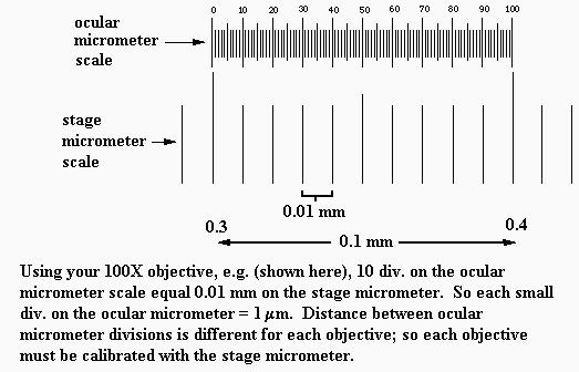
The main purpose of ocular micrometer is to measuer the size of microorganism. The ocular micrometer consists of 2 main scales that are stage scale and ocular scales.

To use this micrometer, we must loacte the ocular scale at the out microscope eyepiece to allow for measurements of objects being viewed. The other scale called stage scales locate at the special slide that contain scales.


We use ocular micrometer to measure the bacterial cell under different magnification.
Discussion
An ocular micrometer is a glass disk that attaches to a microscope's eyepiece. An ocular micrometer has a ruler that allows the user to measure the size of magnified objects. The distance between the marks on the ruler depends upon the degree of magnification. The ruler on a typical ocular micrometer has between 50 to 100 individual marks, is 2 mm long and has a distance of 0.01 mm between marks.

An ocular micrometer is a glass disk that attaches to a microscope's eyepiece. An ocular micrometer has a ruler that allows the user to measure the size of magnified objects. The distance between the marks on the ruler depends upon the degree of magnification. The ruler on a typical ocular micrometer has between 50 to 100 individual marks, is 2 mm long and has a distance of 0.01 mm between marks.
 |
| ocular micrometer |
How to use a ocular micrometer
1. Measure the actual size of the letter on the microscope slide using the millimeter ruler. This measurement will help you calibrate the ocular micrometer to determine if it is giving you accurate measurements.
2. Attach the ocular micrometer to the microscope eyepiece by unscrewing the eyepiece cap, placing the ocular micrometer over the lens and screwing the eyepiece cap back into place. Some microscopes may have an ocular micrometer pre-installed, allowing you to skip this step
3. Slide the stage micrometer onto the microscope slide stage. Adjust the microscope to the lowest possible magnification, which should bring the grid on the stage micrometer into focus.

Move the stage micrometer until the measurement marks on the ocular micrometer align with the measurement marks on the stage micrometer. The measurement "0" on the ocular micrometer should line up with the measurement "0.0" on the stage micrometer.
5. Count the number of measurement marks until the measurements of both the micrometers line up again. At 4x magnification (the lowest setting on most microscopes), the two micrometers will line up again at "3" on the ocular micrometer and "0.3" on the stage micrometer.
6. Write down the number of measurement marks between the aligning measurements for the two micrometers. The distance between measurement marks is 0.01 mm, so you can now determine the distance between coinciding measurement marks. Repeat the exercise at higher magnifications (10x, 40x and 100x), and record these values as well.
7. Use the calibrated ocular micrometer to measure the dimensions of the letter printed on your slide. Compare the dimensions to the dimensions you measured with the millimeter ruler to ensure that the ocular micrometer is functioning properly.
Before using an ocular micrometer, we must calibrated it first. A typical scale consists of 50 - 100 divisions. You may have to adjust the focus of your eyepiece in order to make the scale as sharp as possible. If you do that, also adjust the other eyepiece to match the focus. Any ocular scale must be calibrated, using a device called a stage micrometer.A stage micrometer is simply a microscope slide with a scale etched on the surface. A typical micrometer scale is 2 mm long and at least part of it should be etched with divisions of 0.01 mm (10 µm).
Suppose that a stage micrometer scale has divisions that are equal to 0.1 mm, which is 100 micrometers (µm). Suppose that the scale is lined up with the ocular scale, and at 100x it is observed that each micrometer division covers the same distance as 10 ocular divisions. Then one ocular division (smallest increment on the scale) = 10 µm at 100 power. The conversion to other magnifications is accomplished by factoring in the difference in magnification. In the example, the calibration would be 25 µm at 40x, 2.5 µm at 400x, and 1 µm at 1000x.Some stage micrometers are finely divided only at one end. These are particularly useful for determining the diameter of a microscope field. One of the larger divisions is positioned at one edge of the field of view, so that the fine part of the scale ovelaps the opposite side. The field diameter can then be determined to the maximum available precision.
Conclusion
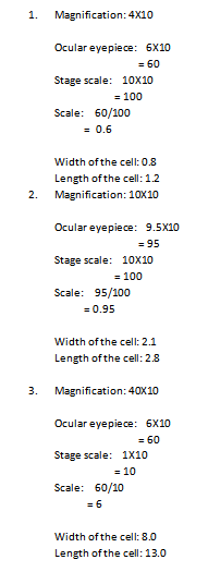
1. This report has identified the correct way to calibrate ocular micrometer. Ocular micrometer has a ruler that allows the user to measure the size of magnified objects. A special slides which contains scales also used to place the objects being observed. Besides, this report also show how to calculate the scale using stage scale and ocular eyepiece. By learning these, small particles such as microorganisms or cell can be measure and the size can be compared.
t
References
http://www.ruf.rice.edu/~bioslabs/methods/microscopy/measuring.html
http://www.ehow.com/how_5019336_use-ocular-micrometer.html
http://www.ehow.com/how_5019336_use-ocular-micrometer.html
Subscribe to:
Comments (Atom)
